- Page d'accueil /
- Livres /
- Medical Books /
- Medicine /
- Surgery /
- Ophthalmology /
- Atlas of OCT: Retinal Anatomy in Health & Pat...
0 ratings

Numéro d'article:
26958971
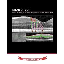
Atlas of OCT: Retinal Anatomy in Health & Pathology Paperback September 30, 2014
Numéro d'article:
26958971
CHF 40
Price Details
Excluding Shipping & Custom charges ( Shipping and custom charges will be calculated on checkout )
*All items will import from États-Unis
0 ratings
Écrire un commentaire
En stock
QTY:
Commandez maintenant et recevez votre commande aux alentours du Jeudi, Juin 25
Transaction sécurisée
Ubuy s'engage à protéger votre sécurité et votre confidentialité. Notre système avancé de sécurité des paiements garantit la confidentialité en chiffrant vos informations lors de la transmission grâce aux protocoles AES (Advanced Encryption Standards) et SSL (Secure Socket Layer). Vos coordonnées de paiement sont 100 % sécurisées car nous ne partageons pas vos informations de paiement avec des vendeurs tiers.
nos meilleurs partenaires logistiques
Become a pro at OCT interpretation with clear and concise color-coded guides.
Livraison
rapide
Retour
gratuit*
Emballage sécurisé
Produits 100 % originaux
Conformité PCI DSS

Certifié ISO 27001
Note: Step Down Voltage Transformer required for using electronics products of États-Unis store (110-120). Recommended power converters Acheter maintenant.


Ce qui se démarque
Comprehensive Retina Guide
This atlas offers extensive insights into retinal anatomy and pathology, catering to both medical professionals and students seeking detailed visual representations of health and disease states.
High-Quality Imaging
Utilizing high-resolution optical coherence tomography (OCT) images, this resource allows for precise observation of retinal structures, greatly enhancing diagnostic capabilities and educational understanding.
Expert Contributions
Authored by leading experts in ophthalmology, this book ensures reliable and authoritative information, making it an indispensable reference for eye care specialists aiming for excellence in patient diagnosis and treatment.
Détails du produit
| Item Weight | 1.4 lbs (640 grams) |
À qui est-ce destiné ?

-
Medical Professionals
Ideal for ophthalmologists, optometrists, and retina specialists seeking detailed insights into retinal anatomy and diseases.
-
Students and Trainees
Beneficial for medical students and residents in ophthalmology, providing foundational knowledge and practical visual reference.
-
Researchers
Useful for researchers studying retinal health and pathologies, aiding in clearer visual interpretations of findings.

-
General Public
Not suitable for laypersons without a background in medical or ophthalmic knowledge who may find it complex.
-
Non-Ophthalmic Specialists
Limited usefulness for healthcare professionals outside of ophthalmology, as the content is highly specialized.
-
Casual Readers
Not recommended for casual readers seeking light reading material, as it focuses on technical medical content.
DESCRIPTION DU PRODUIT
Atlas of OCT: Retinal Anatomy in Health & Pathology Paperback September 30, 2014
Vous avez une question ? Chattez avec nous
Questions et réponses des clients
-
question:
What is the Atlas of OCT: Retinal Anatomy in Health & Pathology about?
répondre: The Atlas of OCT: Retinal Anatomy in Health & Pathology is a comprehensive reference that provides detailed insights into the structure of the retina through Optical Coherence Tomography (OCT) imaging. This atlas covers normal anatomical variations and changes associated with various retinal pathologies. It serves as a crucial resource for ophthalmologists and optometrists to enhance their understanding of retinal diseases and improve diagnostic accuracy. By visually exploring retinal conditions through high-quality OCT images, practitioners can better identify and treat ocular disorders. -
question:
Who is the intended audience for this atlas?
répondre: This atlas is primarily designed for eye care professionals, including ophthalmologists, optometrists, and retinal specialists. It is also beneficial for medical students and residents specializing in ophthalmology, as it provides a solid foundation of knowledge regarding retinal anatomy and pathology. The detailed illustrations and explanations make it a valuable educational tool, enhancing the understanding of OCT imaging techniques for those in clinical practice or training. -
question:
How can the Atlas of OCT be utilized in clinical practice?
répondre: Clinicians can use the Atlas of OCT as a visual guide during patient examinations and diagnosis. By referencing the atlas, they can compare their OCT images with the detailed illustrations of normal and pathological findings. This enhances their ability to recognize retinal conditions accurately, leading to improved patient outcomes. It can also serve as a teaching tool in medical training programs, helping to bridge the gap between theoretical knowledge and practical application in the clinical setting. -
question:
What makes this atlas different from other retinal anatomy books?
répondre: The Atlas of OCT sets itself apart by focusing specifically on Optical Coherence Tomography imaging, which is a cutting-edge diagnostic tool in ophthalmology. Unlike traditional anatomy books, this atlas emphasizes the visual representation of retinal structures as seen through OCT, providing a unique perspective that enhances understanding. Its detailed images paired with concise explanations allow for a more intuitive grasp of complex retinal conditions, making it an indispensable resource for modern eye care practitioners. -
question:
Does the atlas include recent advancements in OCT technology?
répondre: Yes, the Atlas of OCT includes the latest advancements in OCT technology, highlighting the evolution of imaging modalities and their applications in evaluating retinal health. The book discusses new techniques such as swept-source OCT, which offers deeper tissue penetration and improved image resolution. This focus on modern technology allows practitioners to stay updated on the advancements that can enhance their diagnostic capabilities and improve patient care. -
question:
Is the Atlas suitable for beginners in ophthalmology?
répondre: Absolutely! The Atlas of OCT is designed to be accessible even for beginners in the field of ophthalmology. It presents complex concepts in a straightforward manner, supplemented with high-quality images that illustrate key points. Beginners can build a foundational understanding of retinal anatomy and pathology, while also gaining insights into the practical application of OCT. This makes it an excellent starting point for medical students and newly graduated optometrists. -
question:
Can the Atlas of OCT assist in research related to retinal conditions?
répondre: Yes, the atlas can be a valuable resource for researchers focusing on retinal conditions. The comprehensive insights and detailed imaging provided can help in formulating hypotheses, understanding disease mechanisms, and contributing to clinical studies. Researchers can use the images as references to compare their findings, thereby enhancing the quality of their work. This atlas acts as a bridge between clinical practice and research, providing a strong basis for further exploration of retinal diseases. -
question:
What type of images are included in the atlas?
répondre: The Atlas of OCT features a wide variety of images, including high-resolution OCT scans depicting different layers of the retina in both healthy and pathological states. These images illustrate common conditions such as diabetic retinopathy and macular degeneration, alongside normal anatomical structures to showcase the variations. The clarity and detail of these images are crucial for effective learning and understanding of OCT technology, making it an ideal reference for both teaching and clinical practice. -
question:
Are there references for further reading in the Atlas?
répondre: Yes, the Atlas of OCT includes references and suggested readings for further exploration into retinal anatomy and pathology. These references are curated to guide professionals and students towards additional resources that delve deeper into specific topics covered in the atlas. This feature encourages continued education and supports the learning process, enabling users to enhance their knowledge and stay updated with current research findings in the field of ophthalmology. -
question:
Where can I buy Atlas of OCT: Retinal Anatomy in Health & Pathology in Switzerland?
répondre: You can purchase the Atlas of OCT: Retinal Anatomy in Health & Pathology on Ubuy. Ubuy specializes in providing a diverse selection of books and educational resources for healthcare professionals, making it a reliable source for accessing this essential atlas. By shopping on Ubuy, you can ensure that you receive the book conveniently and efficiently, allowing you to enhance your understanding of retinal anatomy and pathology.
Ophthalmology Editorial Review
The Atlas of OCT: Retinal Anatomy in Health & Pathology is a comprehensive guide to the anatomy of the retina using Optical Coherence Tomography (OCT). The book provides detailed images and descriptions of retinal anatomy in both healthy and pathological conditions. It is a valuable resource for ophthalmologists, optometrists, and other eye care professionals.
Avis et évaluations clients
1 évaluations des clients
-
5 étoile
0%
-
4 étoile
100%
-
3 étoile
0%
-
2 étoile
0%
-
1 étoile
0%
Donnez votre avis sur ce produit
Partagez votre avis avec d'autres clients
Avantages
- Comprehensive guide to retinal anatomy
- Detailed images and descriptions
- Useful for ophthalmologists and other eye care professionals
Historique des prix du produit
Informations importantes
- Limitations : Pour les produits expédiés à l'international, veuillez noter que toute garantie du fabricant peut ne pas être valide ; les options de service du fabricant peuvent ne pas être disponibles ; les manuels, instructions et avertissements de sécurité des produits peuvent ne pas être dans les langues du pays de destination ; les produits (et les matériaux qui les accompagnent) peuvent ne pas être conçus conformément aux normes, spécifications et exigences d'étiquetage du pays de destination ; et les produits peuvent ne pas être conformes à la tension et aux autres normes électriques du pays de destination (nécessitant l'utilisation d'un adaptateur ou d'un convertisseur le cas échéant). Il incombe au destinataire de s'assurer que le produit peut être importé légalement dans le pays de destination. En cas de commande auprès d'Ubuy ou de ses filiales, le destinataire est l'importateur officiel et doit se conformer à toutes les lois et réglementations du pays de destination.
- Tous les produits listés sur Ubuy ne sont pas à vendre, Ubuy étant un moteur de recherche mondial. Les produits sont soumis aux réglementations en matière d'exportation et de commerce.
CHF 40
Commandez maintenant et recevez votre commande aux alentours du Jeudi, Juin 25
This item is not restrict in my country.(Please click on above link if this item is not restrict in your country, So our team will review and allow.)
QTY:
Transaction sécurisée
Ubuy s'engage à protéger votre sécurité et votre confidentialité. Notre système avancé de sécurité des paiements garantit la confidentialité en chiffrant vos informations lors de la transmission grâce aux protocoles AES (Advanced Encryption Standards) et SSL (Secure Socket Layer). Vos coordonnées de paiement sont 100 % sécurisées car nous ne partageons pas vos informations de paiement avec des vendeurs tiers.
nos meilleurs partenaires logistiques
Livraison transfrontalière la plus rapide
Caractéristiques et avantages
- Easy interpretation of optical coherence tomography (OCT) images.
- Simple color-coded guides for clear understanding.
- Guides reading steps for interpreting retina and macula images.
- A valuable resource for understanding retinal anatomy in health and pathology.
- A must-have for professionals working in ophthalmology.
- Enhances expertise in interpreting OCT images.